

Free Skin Anterolateral Thigh Flap and Vacuum-Assisted Closure Therapy for Reconstruction of Snakebite-Induced Complex Hand Defects
Introduction
Cobra (Naja species) envenomation remains a serious clinical challenge in tropical regions such as Vietnam. Although systemic effects are often limited, local tissue destruction can be profound, particularly when the hand is involved. Venom-induced cytotoxicity, compounded by secondary infection, frequently results in extensive soft-tissue necrosis affecting skin, tendons, joints, and even bone. These injuries demand aggressive debridement, leaving behind complex defects that are difficult to reconstruct using conventional techniques. This blog discusses a staged reconstructive strategy combining vacuum-assisted closure (VAC) therapy with free anterolateral thigh (ALT) flap transfer to restore form and function in snakebite-induced complex hand defects.
Background and Rationale
Snakebite envenomation is a major public health concern in Vietnam, with cobra bites accounting for a large proportion of cases. The hand is one of the most commonly affected sites, and the extent of local damage often evolves over several days. Cytotoxins in cobra venom cause progressive necrosis of skin, subcutaneous tissue, and fascia, while secondary bacterial infection further worsens tissue loss. Traditional options such as skin grafts or local pedicled flaps are frequently inadequate due to poor vascularity and exposure of vital structures. Therefore, a method that optimizes the wound environment before providing robust, well-vascularized tissue coverage is essential.
Study Overview
This clinical series evaluated a staged approach using VAC therapy followed by free ALT flap reconstruction in patients with complex hand defects caused by cobra envenomation. Eleven patients with extensive skin and/or tendon loss were included. All underwent initial surgical debridement and antivenom therapy, followed by VAC therapy to prepare the wound bed. Definitive reconstruction was then performed using tailored ALT flaps, with postoperative assessment focusing on flap survival, range of motion, and complications.
Patient Selection and Methods
Between January 2020 and April 2025, 214 patients were admitted with cobra bites to the hand. Eleven patients with extensive defects requiring microsurgical reconstruction were managed using the staged protocol. The mean patient age was 39.5 years, with a predominance of male patients. All received antivenom on admission, broad-spectrum antibiotics, and early surgical debridement. VAC therapy was applied for one to two weeks to control infection, reduce edema, and promote granulation tissue before definitive coverage.
Surgical Technique
Initial management involved aggressive excision of all necrotic tissue. A VAC system was then applied at a continuous negative pressure of 125 mmHg, with dressing changes every three to four days. Once a clean, well-granulated wound bed was achieved, reconstruction proceeded using a free ALT flap harvested from the contralateral thigh. Perforators from the lateral circumflex femoral artery were identified, and the flap was designed according to defect size and contour. Depending on the reconstructive requirement, the flap was used as a fasciocutaneous flap or underwent primary or microdissection thinning. The radial artery and accompanying veins were most commonly used as recipient vessels. Postoperatively, the hand was immobilized, followed by gradual initiation of passive and active range-of-motion exercises.
Results
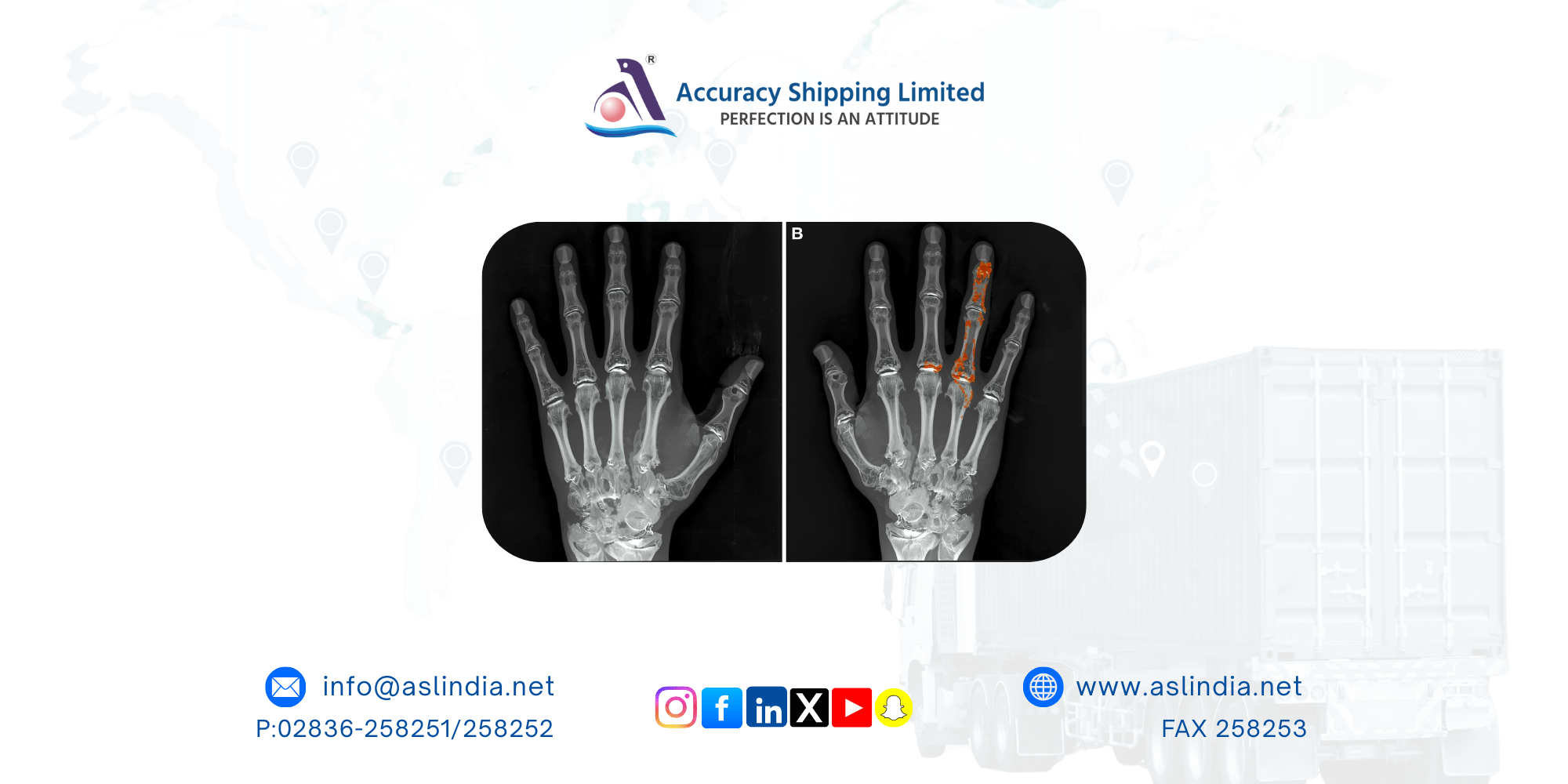
The average duration of VAC therapy was 11.5 ± 4.2 days. All patients demonstrated significant tissue necrosis within 48 hours of the bite, and most had positive wound cultures, with Enterococcus faecalis being the most common organism. Defects frequently involved multiple hand subunits, with exposed tendons or bone in several cases.
A total of 11 free ALT flaps were transferred, with an average flap size of 14.5 × 7.0 cm. Flap thinning was performed in 72.7% of cases, reducing the mean thickness to 4.6 mm. All flaps survived completely, and no donor-site morbidity was observed. Over a mean follow-up of 15.3 months, patients with digital reconstruction achieved functional metacarpophalangeal joint motion, reaching full extension and up to 60 degrees of flexion. The most common postoperative concern was flap bulkiness, but no major complications occurred.
Discussion
Reconstruction of snakebite-induced hand defects is particularly challenging due to progressive tissue necrosis, infection, and compromised local vasculature. Early and thorough debridement is essential to limit toxin spread, but it often leaves large composite defects. VAC therapy plays a critical role in this context by reducing bacterial load, controlling edema, and promoting granulation tissue, thereby creating optimal conditions for microsurgical reconstruction.
The free ALT flap proved highly effective due to its versatility, reliable vascular anatomy, and ability to provide large volumes of well-vascularized tissue. Selective thinning allowed the flap to be adapted for the dorsum and digits, while preserving sufficient bulk for pressure-bearing areas such as the palm. The long vascular pedicle was particularly advantageous, enabling anastomosis outside the zone of venom-induced vascular damage. While musculoskeletal sequelae such as stiffness and tendon adhesions limited complete functional recovery in some cases, the staged approach successfully preserved hand integrity and meaningful function.
Limitations
The study’s limitations include the small sample size and the absence of a control group for comparison with other reconstructive methods. Additionally, although follow-up extended beyond one year in most patients, longer-term assessment is needed to fully evaluate fine motor recovery and long-term functional outcomes.
Conclusion
A staged protocol combining VAC therapy with free anterolateral thigh flap reconstruction offers a safe, reliable, and effective solution for managing complex hand defects caused by cobra envenomation. VAC therapy optimizes the wound environment, while the ALT flap provides adaptable, well-vascularized tissue for multi-component reconstruction. This approach supports durable coverage, promotes functional recovery, and represents a valuable strategy in the management of severe snakebite-related hand injuries.
Our Tag:
Share:
Newsletters
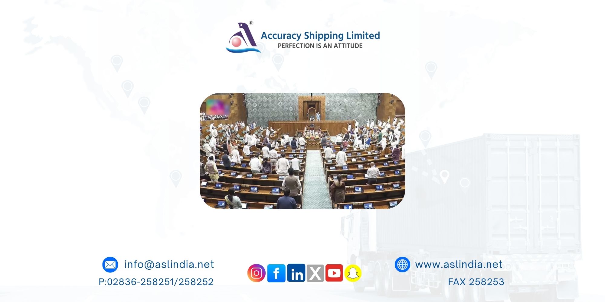
Rafale, Trump, ‘new normal’ with Pakistan: Who said what in Operation Sindoor debate in Lok Sabha

Breakout stocks to buy or sell: Sumeet Bagadia recommends five shares to buy today 12 November 2025

Kolkata port charts independent ship repair strategy

DPIIT partners with YourStory to boost entrepreneurship